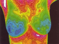
Offered here, is a brief timeline of the development and growing application of thermography in regards to its use as a breast health screening tool. **The following information is based largely on information provided by William C. Amalu of the International Academy of Clinical Thermology, Author of “A Review of Breast Thermography”
Early 1950’s – The use of infrared monitoring systems for night vision by the United States military leads to thermal technology being implemented as a thermal diagnostic tool in medicine.
1957 — Dr. R. Lawson, through the first used of thermographic diagnosis, discovered that the temperature of skin over cancer in breast tissue was higher than that of non-cancerous breast tissue. This leads to thermography being utilized more and more as a breast health screening tool.
1972 — After almost 20 years of trials and use by physicians The Department of Health Education and Welfare took the official stance that thermography would be a useful diagnostic procedure in the area of female breast pathology.
1982 — The FDA publishes its approval of diagnostic thermography in its use as a supplement to traditional breast cancer diagnostic methods.
1986 — An extensive study by H. Useki in breast thermography showed that thermography held an 88% sensitivity level in the detection of breast cancer.
1996 — Dr. P Gamagami writes in his textbook, “Atlas of Mammography” that using thermography as a tool to track angiogenesis – the formation of new blood vessels from pre-existing blood vessels – was successful 86% of the time in detecting early signs of breast cancers that were not otherwise detectable by physical examination and touch alone. He also noted in 15% of these specific cases thermographic imaging helped to show signs of breast cancer where mammography had not.
Since then thermography has been successfully utilized as an adjunct screening tool in many areas including the detection of early signs of breast disorders. Physicians like Dr. Philip Getson of Thermographic Diagnostic Imaging in New Jersey use thermography to help their patients utilize all of the methods they have available in determining risk for breast abnormalities and in the earliest possible detection.
For more information on thermography and the use of thermographic imaging please visit our website here.

