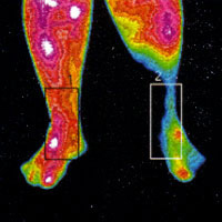
How Neuromuscular Thermography Supports Pain Evaluation
Pain is real, but it rarely shows up on film. X-rays and MRIs image structure, and EMG measures motor nerve function – yet many pain conditions live in physiology those tests don’t capture: autonomic activity, vasomotor changes, the way circulation behaves in an affected limb. Thermography images exactly that layer. It is why pain evaluation has been central to TDI’s work since 1982.
A Physician’s Discipline Behind Every Study
TDI was founded by Philip Getson, D.O., a New Jersey physician who spent decades evaluating patients with complex pain conditions – including Reflex Sympathetic Dystrophy/Complex Regional Pain Syndrome (RSD/CRPS) – and lecturing on the subject at national and international pain conferences. That clinical heritage shaped our pain protocols: standardized bilateral views, controlled room conditions, and marked regions of interest so temperature differences can be measured precisely and reproduced study after study.
What Pain-Pattern Thermography Documents
Side-to-side temperature asymmetry, objectively measured. Skin temperature asymmetry between an affected and unaffected limb is one of the physiological signs clinicians consider when evaluating conditions like CRPS. A thermogram documents that asymmetry visually and numerically – a concrete data point your treating physician can weigh alongside history, examination, and their diagnostic criteria.
Vascular and autonomic patterning. Heat distribution reflects circulation and autonomic activity, which is why thermal images of a painful region often show what the patient has been feeling all along – a limb running colder, a region running hot – in a form that can be seen, measured, and tracked.
A baseline for the long haul. Chronic pain is a long story. Serial thermograms give you and your clinicians a same-protocol record of how your patterns change across months and years.
To be clear about the boundaries: a thermogram does not by itself diagnose CRPS, nerve injury, thoracic outlet syndrome, or any pain condition, and examination, MRI, CT, EMG, and your physician’s judgment remain the backbone of any workup. Diagnosis belongs to your treating clinician. Our job is to hand them – and you – physiological documentation worth having.
The Appointment
Non-contact, unhurried, and private: you acclimate to the temperature-controlled room, the technician captures standardized views of the relevant regions, and a Board-Certified Clinical Thermologist reviews the study. Your written report arrives by email, typically within 2-3 weeks, with an optional phone consultation.
Call 856-596-5834 to discuss which regions to image, or book online.